High Eye Pressure: What to Eat to Lower It?
Nutrition can prove to be a precious ally for vision. So, what to put on the plate when intraocular pressure is high? Among the ingredients to prefer are sources of Omega-3 and carotenoids. Two, in particular, serve the eyes: lutein and zeaxanthin. Let's discover their main sources.
Intraocular pressure (or, more precisely, intraocular) is the pressure of the fluid inside your eye. You can indeed imagine this organ exactly as if it were a balloon filled with water: if it were not full enough, it would collapse; an adequate pressure from inside outwards, exerted precisely by the water, instead keeps it in shape. However, this pressure must not be excessive, under penalty of risk of damage that can seriously compromise vision.
The limit not to be exceeded is 21 mmHg; otherwise, the eye may develop a form of glaucoma – which is one of the leading causes of blindness worldwide. But to diagnose the disease, high intraocular pressure alone is not enough: in the absence of damage to the optic nerve (which transmits nerve signals from the retina to the brain) and signs of glaucoma or other eye diseases, it is simply called ocular hypertension.
Although not a disease, ocular hypertension should not be neglected, even if it affects only one eye. But how to lower intraocular pressure? Are there natural remedies to lower high intraocular pressure or are drugs the only allies to keep it under control? Is there, for example, a link between intraocular pressure and diet that can be exploited to limit the risks associated with possible increases above 21 mmHg?
High intraocular pressure: what causes it??
Precisely because an increase can seriously damage the structures inside the eye, intraocular pressure is carefully regulated by a fluid drainage system that keeps it within acceptable levels (normal intraocular pressure is between 10 and 21 mmHg). However, it may happen that there is an imbalance between the production and drainage of the fluid in the eye and this can increase the pressure inside it – just as it would happen inside a balloon in which water is added.
This fluid is produced by cells located in the back of the anterior eye; after accumulating in the posterior chamber, it flows towards the anterior chamber passing through the pupil. From the anterior chamber, it then flows into veins that drain it outside the eye, while only a small amount returns to the posterior chamber passing through the iris. Its secretion is regulated by the sympathetic nervous system; it is then the intraocular pressure itself that stimulates phenomena that ultimately lead to the drainage of the fluid outside the eye.
So, what influences intraocular pressure: first of all, the proper drainage of the fluid present in the eye. It is also known that intraocular pressure increases slowly with age; for this reason, the risk of ocular hypertension increases after the age of 40. Other risk factors for ocular hypertension and its most feared complication (which, as mentioned, is glaucoma) are high blood pressure (hypertension) and, at the same time, too low blood pressure (hypotension), diabetes, severe myopia, a thin cornea, bleeding at the optic nerve level, trauma or eye surgery, prolonged steroid use, some specific diseases (pigment dispersion syndrome and pseudoexfoliation) and stress.
When is high intraocular pressure dangerous?
Cases of early-onset ocular hypertension are particularly worrying, because an eye exposed to high pressure for many years has a higher probability of being damaged. However, increases should not be taken lightly at any age. Indeed, a sudden increase in intraocular pressure can always stress the fibers of the optic nerve. If this increase persists constantly, intraocular pressure can contribute to the development of so-called primary open-angle glaucoma.
This is a chronic degeneration of the optic nerve. Its connection with ocular hypertension may depend on mechanical damage or the alteration of cells and blood vessels associated with it, which seem to be promoted precisely by the increased pressure inside the eye. For its part, the increase in intraocular pressure leading to glaucoma appears to be associated with mitochondrial dysfunction (organelles within the cell responsible for energy production) and oxidative stress.
What are the symptoms of high intraocular pressure?
Primary open-angle glaucoma is a progressive disease in which the effects of optic nerve damage may take time to manifest. Moreover, in most cases, high intraocular pressure is not associated with particular symptoms. Only rarely is it possible to feel pain in the eyes when moving or touching them. There is no association between high intraocular pressure and headache or dizziness.
The absence of symptoms should not lead to underestimating the risks of high intraocular pressure. Because glaucoma is a slow and progressive disease, even if the optic nerve is not yet damaged and vision is not yet compromised, it is advisable to undergo the appropriate checks to manage the situation in the best possible way and thus reduce the risk of visual field impairment.
What to do in case of high intraocular pressure?
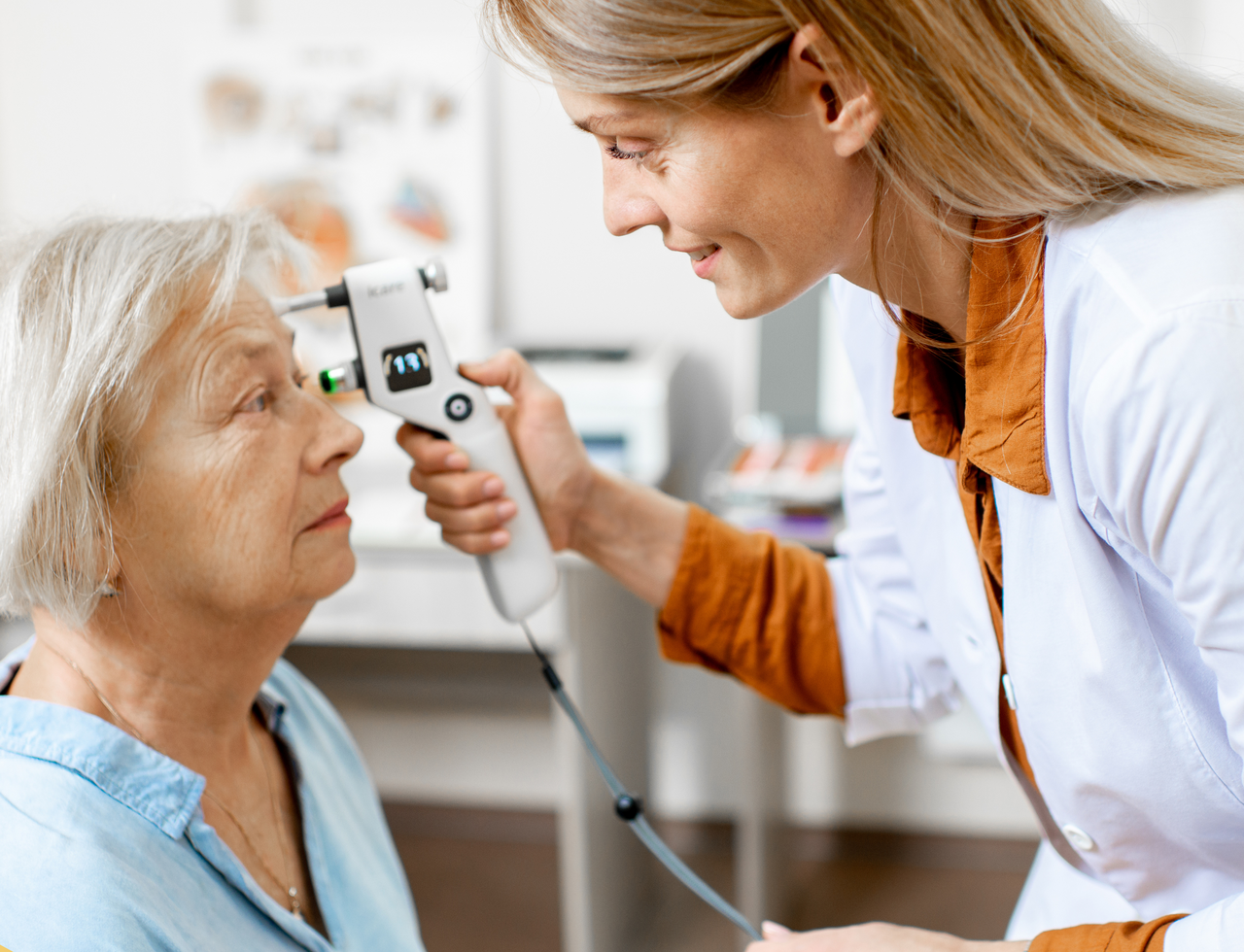
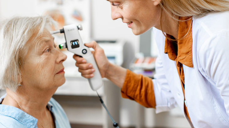
The best approach to managing ocular hypertension is careful monitoring of intraocular pressure, combined with evaluation of the presence of any damage to the optic nerve and other structures and verification of visual abilities. It is therefore essential to undergo all scheduled ophthalmological examinations.
Some cases may require treatment with drugs. The goal is to lower pressure before vision is compromised. The ophthalmologist will assess the need for drug treatment, aiming for personalized management; in particular, the choice to resort to or not to drugs will depend on the risk of developing glaucoma, the presence of signs of optic nerve damage, and the balance between benefits and disadvantages of the treatment.
Surgical treatment is rarer; when needed, laser intervention is generally performed.
What to eat to lower intraocular pressure?
There is no specific diet to lower intraocular pressure. However, a healthy and balanced diet helps protect overall eye health. Among the foods to prefer, fish stands out – a source of Omega-3, allies of vision – and fruits and vegetables, particularly leafy greens (such as kale and spinach), sources of carotenoids that support vision.
The benefits of Omega-3 for vision are certified by Efsa (the European Food Safety Authority), according to which docosahexaenoic acid (DHA, one of the Omega-3s from fish) «contributes to the maintenance of good visual capacities». Not surprisingly, the retina of the eye is rich in DHA. To replenish it at the table, it is advisable to eat fatty fish such as salmon, mackerel, anchovies, sardines, herring, and tuna; in case of increased need, allergies, or a diet that excludes fish for ethical reasons, it can be taken in the form of dietary supplements (remember: there are also sources of DHA suitable for those who cannot or do not want to eat fish).
Regarding carotenoids, those present in food and that end up in the eye are lutein and zeaxanthin, which, unlike the well-known beta-carotene, are not precursors of vitamin A. Per se, lutein and zeaxanthin do not help lower intraocular pressure; however, they seem useful in managing glaucoma due to their marked antioxidant properties.
A review of studies published in the scientific literature in Nutrients in 2021 highlighted the association between a diet rich in carotenoids and the reduction of glaucoma risk; moreover, higher levels of carotenoids in the macula (the central part of the retina, very important for image perception) are associated with better visual capacities in eyes affected by glaucoma. More specifically, lutein has been associated with neuroprotective effects exerted at the retina level.
As mentioned, spinach and kale are rich sources of lutein and zeaxanthin; following a diet richer in these vegetables for at least 4 weeks has been associated with a 4-5% increase in the optical density of the pigment in the macula. Other foods with which to fill up are broccoli, peas, lettuce, egg yolk, parsley, and durum wheat. As in the case of Omega-3, for an additional dose, or to meet increased needs, it is also possible to rely on dietary supplements; their intake, if prolonged for sufficient periods (140 days), has been associated with a significant increase in circulating lutein levels in the body.
Other nutrients allied to vision are the already mentioned vitamin A, abundant in both plant foods (such as carrots and apricots) and animal foods (especially liver), vitamin B12, typically present in animal foods, and zinc, abundant in nuts, meat, and fish. A varied diet that excludes no food groups allows for good intake of these molecules as well. Not only that, a healthy, balanced diet without excess salt helps keep under control some risk factors for ocular hypertension (such as high blood pressure and diabetes).
High intraocular pressure: what to avoid?
To protect eye health, it is instead good to avoid tobacco smoking. In the particular case of ocular hypertension, it is worth reflecting on the link between increased pressure and oxidative stress which, as is known, is promoted precisely by smoking.
Bibliographic references
Abdel-Aal el-SM, Akhtar H, Zaheer K, Ali R. Dietary sources of lutein and zeaxanthin carotenoids and their role in eye health. Nutrients. 2013 Apr 9;5(4):1169-85. doi: 10.3390/nu5041169
Cleveland Clinic. Ocular Hypertension. Last viewed: 30/04/24
European Community. EU Register of Health Claims. Last viewed: 30/04/24
Lem DW, Gierhart DL, Davey PG. Carotenoids in the Management of Glaucoma: A Systematic Review of the Evidence. Nutrients. 2021 Jun 6;13(6):1949. doi: 10.3390/nu13061949
Machiele R, Motlagh M, Patel BC. Intraocular Pressure. [Updated 2022 Jul 11]. In: StatPearls [Internet]. Treasure Island (FL): StatPearls Publishing; 2024 Jan-. Available from: https://www.ncbi.nlm.nih.gov/books/NBK532237/
Salgle G, Montelongo M, Chu l, Mojica S. Carotenoid Supplement Use and IOP Control in Glaucoma Patients. ARVO Annual Meeting Straccar. June 2020. Available from: https://iovs.arvojournals.org/article.aspx?articleid=2768458
